Functional genetic screens are a powerful tool for understanding the genetic underpinnings of biological pathways at a systems level. These screens employ a forward genetics approach, in which cellular phenotypes arising from genome-wide perturbations are analyzed. Causal relationships between the genes and phenotypes are then validated through further screening and experimentation.
There are two modalities of genetic modulation used in screens: gain-of-function or loss-of-function. The former involves driving the expression of a gene so that more mRNA/protein is produced. The latter method involves reducing the amount of or terminating mRNA/protein associated with genes of interest. For both types of manipulation, changes in resulting phenotypes indicate the involvement of a gene in the pathway or disease state of interest.
Over the past decade, new methodological breakthroughs have greatly advanced screening technologies. In particular, the advent of CRISPR (clustered regularly interspaced palindromic repeats) has enabled researchers to deactivate genes in a robust and highly specific manner. This chapter provides an introduction to CRISPR-mediated loss-of-function screens, with a particular focus on identifying and validating gene targets for drug discovery. We will first discuss the role of screens in drug discovery, review CRISPR and earlier technologies used for gene repression, and then outline two formats of CRISPR screens and their components.

The first step of the drug discovery pipeline, called target identification, aims to identify genes (or mRNA or proteins) that are associated with a disease of interest. Loss-of-function screens often play a pivotal role in identifying putative targets. Large-scale primary screens systematically perturb large sets of genes in order to discover targets in an unbiased fashion. For instance, gene disruptions in healthy cells that recapitulate a disease phenotype implicates the gene’s association with the disease. Alternatively, genetic disruptions in diseased cells (e.g., cancer) that cause a normal phenotype can mimic the therapeutic effect of a drug.
In addition to discovering novel drug targets, loss-of-function screens can also be used to improve existing therapies. For instance, one may want to identify genes that confer resistance or increase sensitivity to an existing drug.
Both types of information can be used to make potent drug combinations that are more effective treatments for the disease of interest. Combinatorial screens, in which multiple genes are knocked out, can uncover genetic interactions that can be leveraged for therapy development. Using such screens helps researchers gain insights on the complexities of different diseases and enable the development of more personalized medicines.
Genes identified in the primary screen undergo a rigorous process of target validation to confidently determine whether the identified gene is directly linked to the phenotypic effect. Ways to increase confidence in gene-disease relationships include reproducing the results in biologically relevant cell types, 3D culture, iPS cells, or primary cells taken from patients. Another approach is to design different gRNA sequences for the same gene targets and observe whether the same change in phenotype occurs. Another strategy is to choose an orthogonal method of gene repression. For instance, if RNAi was used in a primary screen, CRISPR can be used in a secondary screen, or vice versa.
Genome-wide functional studies require the ability to specifically target and deactivate a large number of genes in a systematic fashion. Over the past several decades, RNA interference (RNAi) has been a widely used technique to repress genes. Over the past few years, CRISPR-Cas9 technology has emerged as a powerful alternative to RNAi. So what is a CRISPR screen and how does it work?
CRISPR screening is a large-scale experimental approach used to screen a population of mutant cells to discover genes involved in a specific phenotype. Instead of repressing genes at the post-transcriptional level, CRISPR introduces mutations to genes that render them nonfunctional.
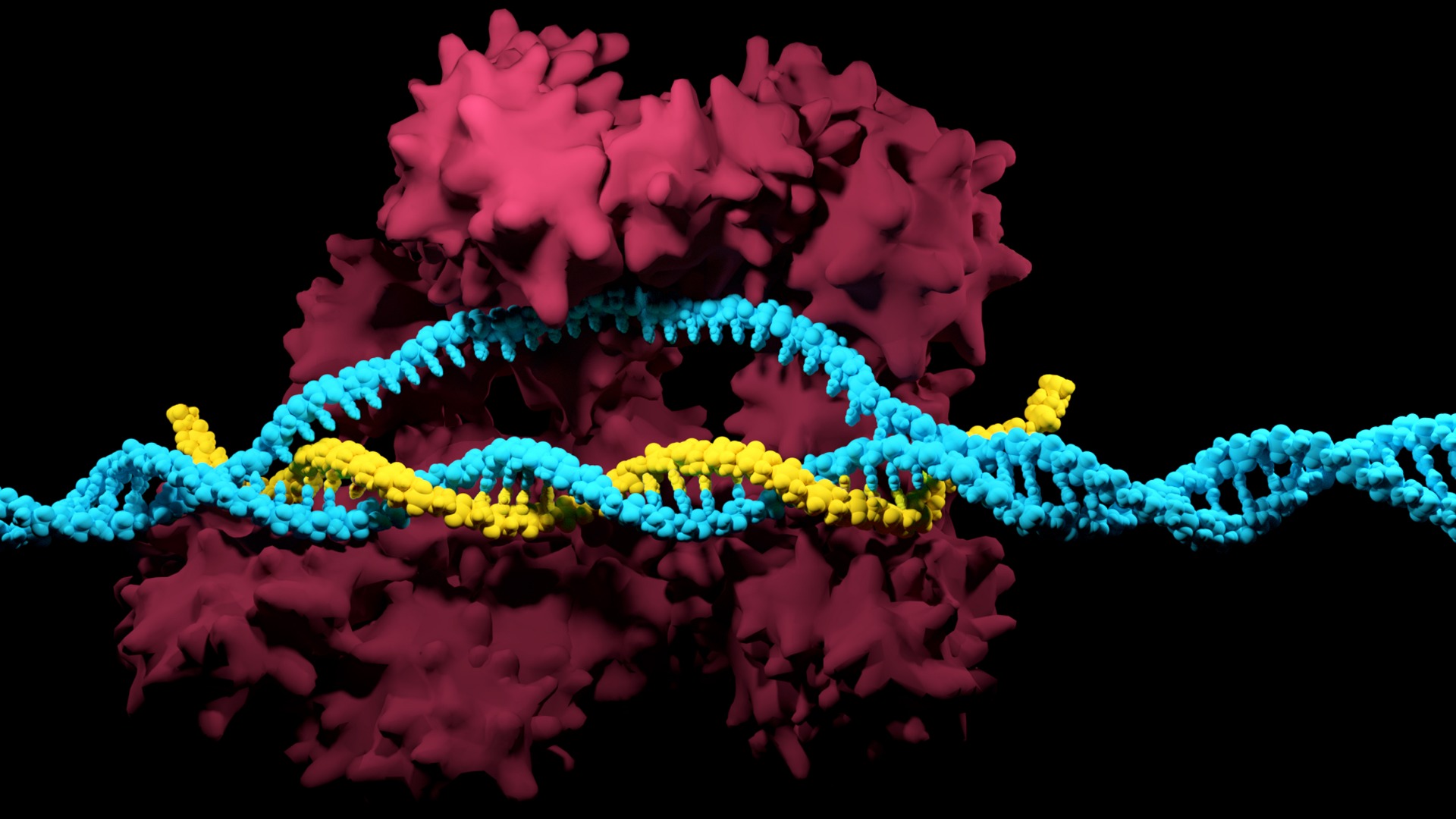
The CRISPR system consists of a programmable guide RNA (gRNA) and a Cas9 nuclease (S. pyogenes) that together form a ribonucleoprotein (RNP). The Cas9 nuclease will make a double-strand break (DSB) in the DNA, thus triggering innate repair mechanisms that can be harnessed for gene editing.
CRISPR-Cas9 has several advantages over RNAi technology (Table 1). Several studies have concluded that CRISPR-Cas9 screens provide more consistent results with fewer off-target effects than RNAi screens (Evers et al. 2016, Shalem et al. 2014, Tan & Martin 2016). Also, because CRISPR-Cas9 editing permanently stops protein expression, it can produce a stronger phenotypic signal and allow for a longer window of analysis. Thus, CRISPR-Cas9 technology is increasingly becoming used in functional genomic screens (Shalem et al. 2015).
Table 1. Benefits and Drawbacks of RNAi and CRISPR

After a set of genes is perturbed, a functional assay is used to qualitatively or quantitatively evaluate the effects of CRISPR-mediated knockouts. Each assay should be customized according to the biological question that is addressed. There are two broad categories of assays: binary and multiparametric.
Binary assays identify cells based on the presence or absence of a desired phenotype. These simple “yes/no” assays can be conducted by either using a selective pressure to kill off a subset of cells (viability assays), or by physically sorting cells into different categories (FACS-based assays).
More sophisticated than their binary counterparts, multiparametric assays measure multiple parameters simultaneously. Thanks to advancements in imaging, microscopy, and other technologies, there are a diverse array of assays that can measure everything from morphological features to the location of proteins in the cell. Some assays can even be used to measure markers of cellular processes over time.
Now that we have explored different types of functional assays, let’s look at how they are applied to CRISPR screening workflows. There are two types of CRISPR screening formats: pooled and arrayed. While both formats can be used to aid in the target identification and validation process, they differ in methodology, equipment, and assay compatibility. Pooled screens involve delivering a mixed population of sgRNA-containing viral constructs into a single tube of cells, while arrayed screens target each gene separately with gRNA across a multiwell plate. Below, we provide an overview of both screen formats.
Pooled screens involve introducing a “pool” of sgRNA into a single population of cells. This is accomplished by packaging sgRNA-containing plasmids into lentiviral particles (one per vector), and then transducing the host cells. The stable expression of guide (along with Cas9) facilities the knockouts of targeted genes. Because knockouts occur across all targets in a single tube of cells, it is difficult to link the phenotype of each individual cell with the underlying genetic perturbation. Pooled screens are thus only compatible with binary assays that physically separate edited cells exhibiting a phenotype of interest from those that do not.
After the cells are sorted, the integrated sgRNAs are sequenced via NGS and data must be deconvoluted. Enriched or depleted sgRNAs in the population confer information about the involvement of corresponding genes in the phenotype.
The arrayed screening format is a newer technology that is more versatile in both methodology and analysis than pooled screens. The arrayed format involves targeting one gene per well across a multiwell plate format. Library delivery may be accomplished through transfection or transduction. Because gene targets are separated across wells, phenotypes do not need to be selected for and sequencing/ data deconvolution are not required to associate phenotypes with genotypes. Arrayed screens are compatible with both binary and multiparametric assays.
The decision on what screening format to use is based on a number of considerations, including the assay, cell model, and labor, equipment availability, and financial costs. A comprehensive comparison of the benefits and drawbacks of each screen format is summarized in Table.
Table 2. Benefits and drawbacks of pooled and arrayed CRISPR screens.

It is important to remember that pooled and arrayed screens can both be useful in screening workflows. For instance, if one aims to identify new drug targets, a pooled format may be appropriate as a primary screen to identify a broad set of target genes in an easy-to-transfect cell model (e.g., immortalized cell line). An arrayed format may then be used in a secondary screen to validate the hits using a more realistic model (e.g., primary cells). When designing a target identification experiment, consider all the tools at your disposal.
As mentioned earlier, having reliable results early in the drug discovery process is critical to reducing the financial risk associated with therapeutic development. The gRNA design in a library can have a profound effect on the outcome of a screen.
For instance, libraries that do not robustly knock out targets may not produce enough detectable signal, and hence lead to false negatives. Additionally, gRNAs that have off-target editing can introduce noise in the system and ultimately complicate untangling or elucidating the causal relationships between genotypes and phenotypes.
Several factors can be taken into account to increase the robustness and specificity of gRNA libraries. For instance, guides can target an early exon or protein-coding genes and be evaluated in silico to minimize off-target editing. Different library designs may also affect efficacy. Libraries from several vendors are designed to rely on individual guides to generate indels. However, because some indels are a multiple of three nucleotides (and do not shift the reading frame), the associated proteins may retain their function. Thus, knockout efficiencies of these libraries may vary, with some genes completely ablated while others are not.
CRISPR has proved to be a compelling tool for functional genomics, as it enables researchers to associate changes in phenotypes with highly specific knockouts. CRISPR-mediated loss-of-function screens are now commonplace for elucidating drug targets for therapeutic development. Two screen types- pooled and arrayed- differ in both methodology and versatility. Whereas pooled screens rely on viral transduction and are limited to assessing binary phenotypes, arrayed screens can be used with a variety of delivery formats and can evaluate complex cellular phenotypes. This latter method is becoming increasingly popular due to the richness of biological information that can be attained.
This protocol describes the use of limiting dilution to isolate single cells (clones) from a CRISPR edited population, the expansion of clonal populations, and screening to identify clones with the...
Thank you for choosing SpCas9 nuclease protein and gRNAs for your CRISPR experiment! SpCas9 has been used for diverse genome editing applications, making it the gold standard in CRISPR gene...
Learn more about clinical applications of CRISPR and how Synthego can support you from early-phase research, through process development, and into the clinic, highlighting our new sgRNA GMP manufacturing capabilities.