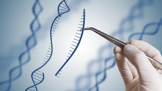
Our understanding of genetics has evolved over the years because of the development of new biotechnologies allowing us to manipulate DNA. CRISPR is one such tool that has soared in popularity because of its ability to precisely cut DNA at a desired location in a genome. In nature, CRISPR is a part of adaptive bacterial immunity, which was later harnessed in research to edit eukaryotic genomes. Researchers can thus mimic a genetic disease by introducing specific genomic mutations associated with the disease in cells helping scientists and doctors understand how the mutant gene impacts cell growth and survival. By understanding the role of the mutated gene in the cell, potential gene or drug therapies can be developed to treat the human disease.
With so much potential to aid our understanding of diseases, you may wonder what factors do you need to consider when designing a CRISPR-based edit in your gene of interest? Or how does one know the CRISPR-Cas system even worked? Here, we highlight the main considerations.
Target the Right Region in a Gene
One of the most important factors influencing CRISPR edits is the region that your guide RNA targets. In this section, we expand more on the do’s and don’ts.
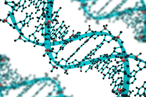
Know Your Introns and ExonsProtein-coding genes are composed of exons and introns. Exons are segments of a gene that code for a protein or peptide. Introns are the gene segments of nucleotide sequences that do not code for a protein. When a protein-coding gene is translated into RNA, introns are removed in a process called RNA splicing. By splicing out introns, the exons come together to be translated into a protein. With most genes containing multiple exons and introns, a cell has found a way to create variants of the same protein from a single gene in a process called alternative splicing. Alternative splicing is a process that involves rearrangement of introns and exons to produce different protein isoforms. Even though these protein isoforms came from the same DNA sequence, they may have different functions in a cell and their expression may be cell-type specific.
Protein-coding genes are composed of exons and introns. Exons are segments of a gene that code for a protein or peptide. Introns are the gene segments of nucleotide sequences that do not code for a protein. When a protein-coding gene is translated into RNA, introns are removed in a process called RNA splicing. By splicing out introns, the exons come together to be translated into a protein. With most genes containing multiple exons and introns, a cell has found a way to create variants of the same protein from a single gene in a process called alternative splicing. Alternative splicing is a process that involves rearrangement of introns and exons to produce different protein isoforms. Even though these protein isoforms came from the same DNA sequence, they may have different functions in a cell and their expression may be cell-type specific.
Beware of IsoformsThe expression of various protein isoforms in a cell can make designing a CRISPR edit experiment difficult. The first dilemma when designing your CRISPR experiments is deciding where you should place the guide RNA in the gene. When attempting to knock out a protein in a cell, it is recommended to design your guide RNA targeting an exon common to most protein-coding isoforms and located at the beginning of the gene to increase the probability that a frameshifting insertion or deletion (indel) of nucleotides could introduce a stop codon. Due to alternative splicing, not all early exons will be present in all protein isoforms. By using online resources such as Ensembl, you can assess if your gene has any isoforms and determine which early exon is present in all isoforms to design your guide RNA over.
The expression of various protein isoforms in a cell can make designing a CRISPR edit experiment difficult. The first dilemma when designing your CRISPR experiments is deciding where you should place the guide RNA in the gene. When attempting to knock out a protein in a cell, it is recommended to design your guide RNA targeting an exon common to most protein-coding isoforms and located at the beginning of the gene to increase the probability that a frameshifting insertion or deletion (indel) of nucleotides could introduce a stop codon. Due to alternative splicing, not all early exons will be present in all protein isoforms. By using online resources such as Ensembl, you can assess if your gene has any isoforms and determine which early exon is present in all isoforms to design your guide RNA over.
Limit Off-Target EffectsOne additional consideration to keep in mind when designing the guide RNA is to limit off-target effects. These off-target effects are unintended indels or mutations caused by the guide RNA directing the Cas nuclease to cut additional regions of the genome other than your targeted gene. By comparing the sequence of your guide RNA with the rest of the genome, you can determine if and how many off-target edits can potentially occur. Synthego’s Guide Validation Tool can help you assess predicted on-target and off-target editing for your designed guide sequence. If you have many potential off-target edit locations, you should reassess the sequence of the guide RNA. Synthego’s CRISPR Design Tool takes these factors into consideration and presents the top suggested guide RNA sequences that fit these parameters.
One additional consideration to keep in mind when designing the guide RNA is to limit off-target effects. These off-target effects are unintended indels or mutations caused by the guide RNA directing the Cas nuclease to cut additional regions of the genome other than your targeted gene. By comparing the sequence of your guide RNA with the rest of the genome, you can determine if and how many off-target edits can potentially occur. Synthego’s Guide Validation Tool can help you assess predicted on-target and off-target editing for your designed guide sequence. If you have many potential off-target edit locations, you should reassess the sequence of the guide RNA. Synthego’s CRISPR Design Tool takes these factors into consideration and presents the top suggested guide RNA sequences that fit these parameters.
Select a Suitable Cell Line
Picking the right sequence of your guides to successfully conduct a CRISPR edit is not the only choice you will have to make. Selecting your cell line is very important because each cell line reacts differently to CRISPR editing. For example, immortalized cells such as HEK293 and HeLa cells are a couple of easy cell lines to work with. On the other hand, primary cells are difficult to work with because it is hard to deliver foreign molecules into them like in transfection procedures. Primary cells are also difficult to work with because they have the ability to drift and change characteristics to match their new growth conditions. Another category of cells that is widely used in research is induced pluripotent stem cells (iPSCs). iPSCs are unique in that they have the ability to transform into any cell type by a process called differentiation. Each of these cell types and their advantages and caveats in research has been explained in more detail in our ‘Stop Sabotaging Your CRISPR Experiment with Wrong Cell Type’ blog post.
Choose an Appropriate Transfection Method
Once you have chosen your cell line for your experiments, you will need to artificially introduce the components of CRISPR, the guide RNA and Cas nuclease, to your cells through a process called transfection. The three common types of transfection are electroporation, lipofection, and nucleofection.
At Synthego, we have designed a protocol for each of these transfection methods for immortalized cell lines:
In most cases, transfection of the CRISPR-Cas complex into your cells will generate a mixed population of cells called a pool consisting of cells with no edits (wildtype), heterozygous edits, or homozygous edits. To create a population of cells that are all clones of each other consisting of one type of CRISPR edit, you will want to isolate a single cell for clonal expansion. Following Synthego’s Limiting Dilution protocol, we walk you through how to properly isolate cells from your pooled cell population to a clonal cell population.
For additional transfection protocols for iPSCs or T cells, please check out Synthego’s protocol webpage.
Confirm the Success of Your CRISPR Edits
Whichever cell line you do choose, it is crucial you validate your CRISPR edits to confirm that your experiments worked. This should be done at the genomic level and protein level, as described below.
Genotype to Identify Sequence ChangesThrough a process called genotyping, you will be able to assess the DNA sequence where your CRISPR edit was predicted to occur. Synthego’s Genotyping Protocol will walk you through the steps to perform Sanger sequencing of your CRISPR edited samples. Using bioinformatics tools, such as Synthego’s Inference of CRISPR Edit (ICE), you will be able to analyze your Sanger sequencing files to validate if your CRISPR experiments successfully altered the DNA sequence. With that said, if your experimental intentions are to alter protein expression, the presence of protein in your samples must be determined.
Through a process called genotyping, you will be able to assess the DNA sequence where your CRISPR edit was predicted to occur. Synthego’s Genotyping Protocol will walk you through the steps to perform Sanger sequencing of your CRISPR edited samples. Using bioinformatics tools, such as Synthego’s Inference of CRISPR Edit (ICE), you will be able to analyze your Sanger sequencing files to validate if your CRISPR experiments successfully altered the DNA sequence. With that said, if your experimental intentions are to alter protein expression, the presence of protein in your samples must be determined.
Assess Protein Depletion with AssaysProtein expression assays allow you to quantify how much of the protein you are studying is present in your cells. One of the gold standard methods for assessing protein expression is ‘western blot analysis’. A western blot requires the binding of a unique antibody to the protein of study to detect the expression levels of that protein. A commonly expressed protein such as glyceraldehyde 3-phosphate dehydrogenase (GAPDH), can be used as a control to compare expression levels of the protein being studied. However, what happens when you see pervasive expression levels of the protein you study? Did your CRISPR experiments even work? What possible reasons could have influenced your CRISPR experiment not to work?
One of the most common reasons why you may observe pervasive protein expression in your knockout cells is that not all isoforms of the protein were considered when designing your guides. For instance, in knockout CRISPR experiments, there should be low to no protein expression of the protein if the edit was successful. If not all of the prominent isoforms were considered when designing your guides, one or more of those isoforms can still be expressed. Additionally, depending on the cell type and target gene, due to alternative start sites and exon skipping phenomena, a truncated form of the protein may still be expressed. These alternative isoforms can still be detected on your western blot analysis and could also still be functional within the cell. A solution is to look back at your initial guide designs and double-check if your guide is placed over an exon that is present in all the prominent isoforms of that transcript. If it is not, it is recommended to redesign your guides to target an early exon, an exon present in all prominent isoforms, and select the guides with minimal off-target effects.
The most important selections for your CRISPR edit experiments include choosing the best region within your gene of interest that creates a successful knockout of the protein while minimizing off-target effects, selecting a viable cell line you are comfortable working with and is relevant to your research, and selecting the best transfection method to ensure successful CRISPR-Cas complex delivery. By selecting the best options outlined above for your CRISPR experiment, you will be on your way to successfully understand how your gene of interest works in a cell.
Protein expression assays allow you to quantify how much of the protein you are studying is present in your cells. One of the gold standard methods for assessing protein expression is ‘western blot analysis’. A western blot requires the binding of a unique antibody to the protein of study to detect the expression levels of that protein. A commonly expressed protein such as glyceraldehyde 3-phosphate dehydrogenase (GAPDH), can be used as a control to compare expression levels of the protein being studied. However, what happens when you see pervasive expression levels of the protein you study? Did your CRISPR experiments even work? What possible reasons could have influenced your CRISPR experiment not to work?
One of the most common reasons why you may observe pervasive protein expression in your knockout cells is that not all isoforms of the protein were considered when designing your guides. For instance, in knockout CRISPR experiments, there should be low to no protein expression of the protein if the edit was successful. If not all of the prominent isoforms were considered when designing your guides, one or more of those isoforms can still be expressed. Additionally, depending on the cell type and target gene, due to alternative start sites and exon skipping phenomena, a truncated form of the protein may still be expressed. These alternative isoforms can still be detected on your western blot analysis and could also still be functional within the cell. A solution is to look back at your initial guide designs and double-check if your guide is placed over an exon that is present in all the prominent isoforms of that transcript. If it is not, it is recommended to redesign your guides to target an early exon, an exon present in all prominent isoforms, and select the guides with minimal off-target effects.
The most important selections for your CRISPR edit experiments include choosing the best region within your gene of interest that creates a successful knockout of the protein while minimizing off-target effects, selecting a viable cell line you are comfortable working with and is relevant to your research, and selecting the best transfection method to ensure successful CRISPR-Cas complex delivery. By selecting the best options outlined above for your CRISPR experiment, you will be on your way to successfully understand how your gene of interest works in a cell.