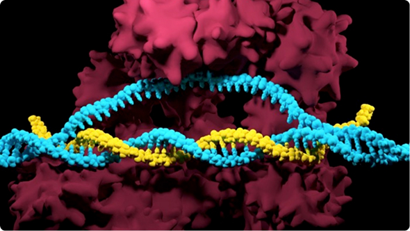
A key step of any CRISPR workflow is delivering the gRNA and Cas9 into the cytoplasm or nucleus of target cells. The delivery of nucleic acids into eukaryotic cells is called transfection. The gRNA and Cas9 can be introduced as either DNA, RNA, or pre-complexed RNA and protein called ribonucleoproteins (RNPs). Transporting foreign materials across multiple cellular barriers (e.g., plasma membrane, nuclear membrane) can be a challenge. Multiple methods have been developed to introduce nucleic acids into cells while maintaining cell viability.Transfection methods can be broadly classified into physical, chemical, and viral-mediated categories. Each method has different advantages and disadvantages with respect to efficiency, throughput, equipment, skill, and cost. In this guide, we discuss some tips to consider before choosing a transfection protocol and review the advantages and disadvantages of each method.
1. Transient vs. Stable Transfection
The method used depends on whether one wants the Cas9 protein and/or the guide RNA to be present temporarily or be permanently expressed in the transfected cells. In a transient transfection, the CRISPR components are introduced into the cell, but no DNA encoding a guide RNA or Cas9 are incorporated into the cell’s genome. CRISPR-Cas9 can only cleave the cell’s genomic DNA for a limited period of time with this approach. One main advantage of transient transfections is that this approach limits the risk of off-target effects because the CRISPR components do not edit the genome over long periods of time.
For certain experiments where long-term expression of gRNAs and/or Cas9 are required, it may be preferable to follow a stable transfection protocol, in which DNA encoding one or both CRISPR components is permanently inserted into the cell’s genome. Because the genes are integrated into the cellular genome, they will be passed down to future generations of cells after cell division. The expression of Cas9 and/or guide RNA genes can be either inducible (e.g., by a trigger, such as a drug) or constitutive (occurring at all times).
Stable transfections are most effectively achieved by using a viral vector, though they can also occur at low frequency following the introduction of plasmid DNA. It is common for researchers to stably transfect only DNA encoding Cas9 (but not guide RNA) to produce a Cas9-expressing cell line. Then, gRNAs for different targets can be introduced to the Cas9-expressing cells through transient transfection.
Stable transfections are generally more laborious than transient transfections. They often involve co-transfecting a selectable marker (e.g., an antibiotic resistance gene), enriching for cells with the successful genomic integration, and cloning of selected cells. Because stable transfections involve more work, they are usually generated only for research projects where long-term expression of CRISPR components is required.
2. Transfection Throughput
Your choice of a CRISPR transfection protocol may depend on the number of samples you need to process at a time. For large numbers of samples, a high-throughput method is preferable. These methods often involve the use of automated equipment or more advanced machinery. Alternatively, a low-throughput method may be adequate for smaller numbers of samples. Some sample types, such as mouse embryos, can only be efficiently transfected using low-throughput methods.
3. Transfection Equipment & Reagents
Several transfection methods require specialized equipment and reagents. The choice of a protocol may largely depend on whether you have access to required equipment or are willing to purchase it. Such decisions may be influenced by your available resources and how much gene editing work you plan to do.
4. Format of CRISPR Components
The format of your CRISPR components may affect your decision of which transfection method to use. Guide RNA and Cas9 can be delivered to cells as DNA, RNA, or as pre-formed ribonucleoprotein complexes (RNPs) formats. The DNA and RNA formats for Cas9 require transcription and/or translation after being introduced into the cell and the the nuclease needs to cross the nuclear envelope after it’s made.
The translocation of DNA/RNA into the nucleus is mediated by the nuclear pore complex and mitosis (when the nuclear envelope breaks down and then re-assembles). If using these formats, one should include a nuclear localization sequence (NLS) in the sequence of Cas9 to ensure it can enter the nucleus for genome editing. As far as choosing a transfection method, one can choose whether they prefer the components be transported to the cytoplasm (electroporation, lipofection) or the nucleus (nucleofection, microinjection).
Alternatively, the pre-formed RNP format does not require any transcription or translation. If using RNPs, then choosing a transfection method that delivers to the nucleus (nucleofection, microinjection) may be favorable as it will enable editing to occur quickly because fewer steps are required.
Summary of CRISPR component delivery options:
- DNA: DNA will enter the nucleus to be transcribed into RNA, either the mRNA that will be translated into Cas9 or the gRNA. The mRNA encoding Cas9 will then enter the cytoplasm to be translated to Cas9 protein. The Cas9 and gRNA then unite in the cytoplasm to form an RNP and move back into the nucleus by crossing the nuclear envelope to target genomic DNA.
- RNA: The Cas9 mRNA will be translated into protein in the cytoplasm. The gRNA and Cas9 then form an RNP in the cytoplasm and enter the nucleus.
- Ribonucleoprotein complex (RNP): Pre-complexed RNPs can be delivered to the cytoplasm or directly to the nucleus without any additional requirement for transcription or translation.
5. Cell Type and Efficiency of Transfection
Transfection efficiency often depends on cell type. Most common cell lines and immortalized cells are generally easy to transfect. Primary cells and stem cells, on the other hand, are more sensitive and often have lower viability after transfection.
Immortalized cell lines
Immortalized cell lines propagate indefinitely in laboratory cell culture conditions. They are either derived artificially by inducing mutations that prevent senescence, or naturally from cancers. Because immortalized cells divide frequently (during which the nuclear envelope is broken down), there are ample opportunities for CRISPR components to enter the nucleus. Thus, these cell lines are generally easy to transfect. One drawback of cell lines is that they can accrue genetic mutations over time, and these changes can be significant.
Because of this, cell lines may be aneuploid and are less representative of the natural biology of living tissues. They are also vulnerable to contamination. However, because they are cost-effective and easy to culture and manipulate, immortalized cells are commonly used in research, especially for early proof-of-concept experiments.
Examples: HEK293, HeLa, A549, Jurkat
Primary cell
Primary cells are derived directly from living tissue without introducing additional genetic changes. Primary cells are subdivided into two types: adherent (requiring attachment to a surface in culture) or suspension (not requiring attachment). The ability of primary cells to propagate is limited and therefore, they cannot be cultured for extended periods of time.
Because primary cells have limited numbers of cell divisions, there are fewer opportunities for CRISPR components to enter the nucleus. Thus, these cells are typically difficult to transfect. However, primary cells may undergo a process called transformation, either naturally or following stimulation, in which they gain mutations that make them immortalized. A significant benefit of primary cells is that they are more representative of the natural biology of living tissues than immortalized cells.
Examples:
- Adherent: cells from organs (kidney, liver, epithelial cells, fibroblast)
- Suspension: cells from blood (T cell, lymphocyte)
Stem cells
Stem cells have natural properties that enable them to propagate in an undifferentiated state for an indefinite amount of time. When cultured in the lab, they can either be maintained as undifferentiated cells or stimulated to differentiate. Stem cells can either be pluripotent (able to differentiate into any cell type) or multipotent (able to differentiate into some cell types). The main types of stem cells are embryonic stem cells (ES cells), adult stem cells, and induced pluripotent stem cells (iPS cells).
Examples: iPS cell, ES cell, Hematopoietic stem cells (HSC)
Germline cells, zygotes, and embryos
Germline cells develop into gametes (sperm and eggs) and can be transfected before or after fertilization. In sexually-reproducing organisms, a sperm fertilizes an egg, producing a single-celled zygote. The zygote then divides and develops into a multicellular embryo. Gametes and embryos are generally difficult to transfect by most methods.
Examples: mouse pronucleus (the nucleus of a fertilized egg), insect eggs, embryos
|
Lipofection |
Electroporation |
Nucleofection |
Microinjection |
Virus |
| Principle |
Lipid complexed with genetic material fuses with the cell membrane |
Electric pulse forms pores in the cell membrane for entry of DNA/RNA/RNP |
A electropotation-based optimized for nuclear deliver Pre-optimized for each cell type |
Microneedle injects CRISPR components inside cells, oocytes, or zygotes |
DNA/RNA is packaged into infectious particles & introduced into cells |
| Advantages |
Cost-effective, High throughput |
Easy, Fast, High efficiency |
Easy, Fast, High efficiency |
High efficiency |
High efficiency |
| Limitations |
Less efficient |
Requires optimization |
Requires reagents & equipment |
Time-consuming, Technically demanding, Low throughput |
Time-consuming, Safety requirements Expensive |
| Cell Types |
Few |
Numerous |
Numerous |
Few |
Numerous |
Physical Transfections of CRISPR Components
Physical transfection protocols create temporary holes in the plasma membrane through which gRNA/Cas9 can pass. Three popular physical methods to introduce CRISPR components into cells are electroporation, nucleofection, and microinjection. Electroporation and nucleofection use an electrical pulse to create pores in the plasma membrane, while microinjection uses a needle to force a hole through the membrane.
Electroporation involves suspending cells in a conductive solution and briefly applying high-voltage electrical pulses. The application of electricity induces the formation of temporary pores in the plasma membrane and the electrical potential across the membrane causes charged molecules (e.g., gRNA/Cas9) to enter the cytoplasm through the pores. Nucleofection is based on electroporation, but utilizes a special machine, called a Nucleofector, and has several distinctions from traditional electroporation (discussed below). Electroporation and nucleofection are effective for transfection of many cell types.
Figure 1. Electroporation/Nucleofection
Traditional electroporation and nucleofection are based on the same overall methodology. Cells are first mixed with an electroporation or nucleofection reagent, and then added to CRISPR components (shown here as RNPs). The cell-RNP mix is then put into a Nucleocuvette (for nucleofection) or Neon pipette tip (for electroporation) and inserted into a Nucleofector or electroporator. Once in the machine, an electric pulse is applied to the cells. The electric field temporarily causes pores to form in the plasma membrane, enabling RNPs to enter the cells. Nucleofection also enables nuclear entry of RNPs. Once the electric field is removed, the plasma membrane is repaired.
Advantages:
- Fast and easy
- High efficiency
- Large numbers of cells can be transfected in a short time (minutes)
Disadvantages:
- Requires specialized equipment
- Cell death may result from high voltage pulses and incomplete membrane repair
Cell Types: immortalized cells, primary cells, stem cells, oocytes/zygotes/embryos
Differences Between Traditional Electroporation & Nucleofection
Electroporation-based methods can be subdivided into two types: traditional electroporation and nucleofection. Traditional electroporation collects the cells, gRNA/Cas9, and reagents in a pipette tip or a specialized electroporation cuvette. The electrical pulse is then applied to the components to facilitate the introduction of the CRISPR components across the plasma membrane, and sometimes also into the nucleus.
There are only a few buffers available that are compatible with electroporation, and each sample must be processed one at a time (low-throughput). A benefit of this device is that it is an open system and the user can manually set the parameters for each sample. This characteristic allows one to optimize specific parameters for each cell type. A popular electroporation device is ThermoFisher Scientific’s Neon Transfection System.
Immortalized Cell Electroporation Protocol
Our CRISPR Neon electroporation protocol was designed to allow highly efficient transfection of RNP complexes into standard cell lines. Achieve high editing efficiencies in your CRISPR experiments with minimal optimization.
Download
Nucleofection uses a Nucleofector (Lonza) machine designed to facilitate the transfer of gRNA/Cas9 directly to the nucleus. The procedure involves combining target cells, a buffer specific to each cell type, and gRNA/Cas9. These components are transferred to a cuvette or 16-well strip, which is then placed into a Nucleofector. An electric pulse with parameters pre-optimized for each cell type is then applied.
The specificity of the solution and electric pulse enables the gRNA/Cas9 to directly enter the nucleus. Because cell division is not required for the transfer into the nucleus, nucleofection is an effective method for non-dividing cells (e.g., neurons, blood cells). Multiple samples can be transfected at once, making it a higher throughput method than traditional electroporation. However, a drawback of the nucleofector is that it is a closed system (.i.e., “black box”). The parameters of each program are not identified and cannot be individually controlled by the user. The components of buffers are also proprietary. The Nucleofector may also be more expensive than other electroporation systems.
Nucleofection with RNPs is the preferred method to introduce the CRISPR components into most cell types in terms of editing efficiency. The use of chemically modified synthetic single guide RNA has been shown to increase editing efficiencies in immortalized, primary, and stem cells.
Immortalized Cell Nucleofection Protocol
Our CRISPR nucleofection protocol was designed to allow highly efficient transfection of RNP complexes into standard cell lines. Achieve high editing efficiencies in your CRISPR experiments with minimal optimization.
Download
Microinjection
Microinjection involves carefully positioning a target cell under a microscope and delivering gRNA/Cas9 into the cytoplasm or nucleus through a glass micropipette. This method is highly technical and requires special equipment, including micromanipulators and a microinjector. It is also low-throughput, as only one cell can be transfected at a time.
However, this method can result in very high efficiency when performed by skilled individuals. Microinjection is mainly used for transfecting single cells, oocytes, zygotes, and embryos. It is a popular method for pronuclear injections (into the nucleus of fertilized eggs), which is used to generate transgenic mice and other animals.
Figure 2. Microinjection
Microinjection utilizes a microscope, microinjector, and other equipment (not shown) to inject CRISPR components (shown here as RNPs) into the cytoplasm or nucleus of cells using a glass microinjection needle. Each cell must be injected individually, making microinjection a low-throughput process.
Advantages:
- High efficiency (near 100%)
Disadvantages:
- Labor-intensive & requires skill
- Damage to the plasma and nuclear membranes can lead to cell death
- Low-throughput
- Requires special equipment
Cell Types: single cells, oocytes, zygotes, and embryos.
Mouse Embryo Microinjection Protocol
This CRISPR mouse embryo microinjection protocol, provided by researchers at the University of Rochester Mouse Genome Editing Resource, was designed to allow highly efficient introduction of synthetic single guide RNA into mouse embryos. Achieve high editing efficiencies in your CRISPR experiments with minimal optimization.
Download Protocol
Chemical Transfections of CRISPR Components
Several chemically-based methods can be used to transport molecules into cells, including calcium phosphate, cationic polymers, and cationic amino acids. One of the most common methods to introduce CRISPR components into cells is lipofection.
Lipofection (also called lipid-based transfection) uses cationic lipid reagents to deliver CRISPR components into cells. This method first involves constructing lipid-soluble structures, called liposomes, around the CRISPR components. The components are then transported into the cell through endocytosis, a process in which the plasma membrane surrounds the components on the exterior-side, and buds off inside the cell. The CRISPR components then escape the endosomal pathway and diffuse through the cytoplasm. Unlike nucleofection and microinjection, lipofection does not deliver CRISPR components to the nucleus.
Lipofection is an easy and economical method that usually has minimal toxicity. However, lipofection efficiency is usually not as high as the other methods This method is not recommended for primary cells or stem cells.
Figure 3. Lipofection
Lipofection utilizes lipids to transport CRISPR components (shown here as RNPs) into cells. The RNPs are mixed with a lipofection reagent containing positively-charged (cationic) lipids. The lipids form vesicles, called liposomes, surrounding the RNPs. When delivered to cells, the liposomes are transported across the plasma membrane through endocytosis. Once inside the cell, the RNPs escape the endosomal pathway and diffuse through the cytoplasm. The CRISPR components can only enter the nucleus during mitosis or as RNPs tagged with a nuclear localization sequence (NLS).
Advantages:
- Economical & easy
- Can be adapted to high-throughput systems
- Low cytotoxicity
- Does not require specialized equipment
Disadvantages:
- Moderately efficient
- Limited cell types
- No delivered directly to the nucleus
Cell Types: Standard cell lines and immortalized cells
Immortalized Cell Lipofection Protocol
Our CRISPR lipofection protocol was designed to allow highly efficient transfection of RNP complexes into standard cell lines without using any specialized equipment. This protocol is best optimized for medium-throughput experiments in 24-well plates.
Download Protocol
Viral Transfection Methods
Viral vectors can be used to transfer DNA or RNA into cells in a process called transduction. The process first involves packaging the gRNA/Cas9 sequences into viral particles and then introducing the particles into target cells. To make the viral particles, the plasmid containing the gRNA or Cas9 sequence and plasmids containing viral genes are introduced into a packaging cell line (e.g., 293 T cells). Once the viral particles are produced, they are harvested from the packaging cells and introduced into the target cells that one wants to transfect with gRNA or Cas9.
There are several types of viruses that can be used for transduction, including lentivirus, adenovirus, adeno-associated virus, and herpes viruses. For stable transduction, lentiviruses, a family of retroviruses that includes HIV, are often used because they integrate their genome into the genome of infected cells (Fig 4). Lentiviruses can penetrate the nuclear envelope, and thus are effective in non-dividing cells (i.e., it is not reliant on mitosis for nuclear entry). Lentiviruses are effective in a variety of cell lines, primary cells, and stem cells, and can be used in vivo. However, because they integrate randomly into the genome, they may disrupt vital genes or alter the regulation of gene expression.
Adeno-associated virus (AAV) is another type of virus commonly used for in vivo editing and often results in the sustained expression of exogenous genetic sequences in dividing and non-dividing cells. AAVs typically do not trigger an immune response and have a range of serotypes that enable targeting to different tissues. Another benefit of recombinant AAVs is that they do not integrate into the genome, and will not cause disruption of genes through insertional mutagenesis.
One drawback of AAVs is their limited packaging capacity of about 4.5 kb, so it may be a challenge to fit both gRNA and Cas9 sequences in a single vector. AAVs are also commonly used to transport donor DNA templates into cells for knock-in experiments.
Viral vectors are highly efficient at transfecting CRISPR components and compatible with many cell types. However, constructing viral particles is time-consuming and laborious. Depending on the type of virus used, extra safety measures may be required in the laboratory.
Figure 4. Lentiviral Transduction
Virus-based transfections are referred to as transductions. Lentiviruses can be used for stable transfections of gRNA/Cas9. The viral particles must first be constructed using a packaging cell line. The gRNA/Cas9 sequences, viral packaging genes, and viral envelope genes are each put on separate plasmids, and then introduced into the packaging cells. The constructed viral particles are then harvested and introduced into the target cell line (transduction). Once the viral particles cross the plasma membrane and the viral RNA is reverse-transcribed into cDNA. The cDNA then enters the nucleus and is randomly incorporated into the genome of the host cell.
Advantages:
- High efficiency
- Compatible with many cell types
- Can be used in vivo and in vitro
Disadvantages:
- May require extra safety measures
- Laborious & time-consuming
- Some types may have immunogenicity & cytotoxicity
What CRISPR Transfection Protocol is Best for You?
There isn’t a “one-size-fits-all” answer to what protocol everyone should use for their CRISPR experiments. The sample type and experimental goals will primarily determine the best protocol for each experiment, and in every case, the protocol selected will require optimizations to achieve the highest editing efficiencies.
However, researchers at Synthego have determined that the best method for introducing CRISPR components into the majority of cell types involves Nucleofection of RNP complexes consisting of chemically modified synthetic sgRNA and purified recombinant Cas9.
Visit the Resources section of our website to check out our complete list of CRISPR protocols and other guides to help you achieve the highest level of success in your genome engineering experiments.
