CRISPR is famous for its gene editing capabilities, making waves in the fields of therapeutics, diagnostics, and agriculture, just to name a few. However, researchers also use CRISPR for an application that has nothing to do with gene editing: visualization of DNA.
Scientists are using the principles of CRISPR and its components to visualize the three-dimensional organization of cellular components in real time. The concept of visualizing cells is referred to as imaging. In this article, we will discuss the field of imaging and highlight the impact CRISPR is having on it by breaking it down into the following sections:
Before we talk about how CRISPR is changing the imaging field, we first need to describe the techniques that are traditionally used by scientists for cell imaging, and their limitations.
There are different methods scientists use to image cells based on 1) what cellular element they want to image (i.e., protein or nucleic acid) and 2) if they need to see it in real-time (i.e., live-cell or fixed). This section describes the various methods, the similarities and differences, and limitations.
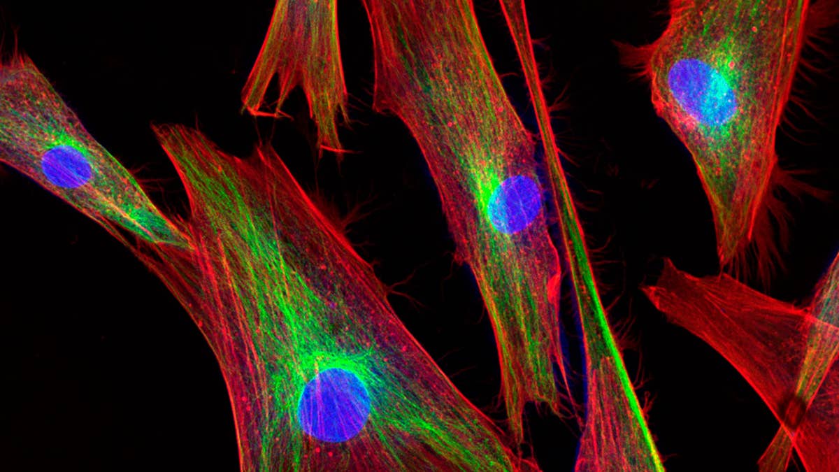
Protein imaging is commonly done using techniques called immunofluorescence (IF) or immunohistochemistry (IHC). These techniques rely on cell fixation, a process that ‘freezes’ the cell in that moment in time (like a snapshot) by killing it. Following fixation, the cells are permeabilized and incubated with antibodies that recognize the protein of interest. Finally, scientists add a detection agent, usually a fluorescent or colorimetric protein that acts as a readout for their target.
Scientists are also able to image live cells (no fixation necessary) by engineering or tagging proteins with fluorescent tags. These fluorescently labeled proteins can then be tracked in real time visually by microscopy. Live-cell imaging works best with abundant proteins and a robust fluorescent signal.
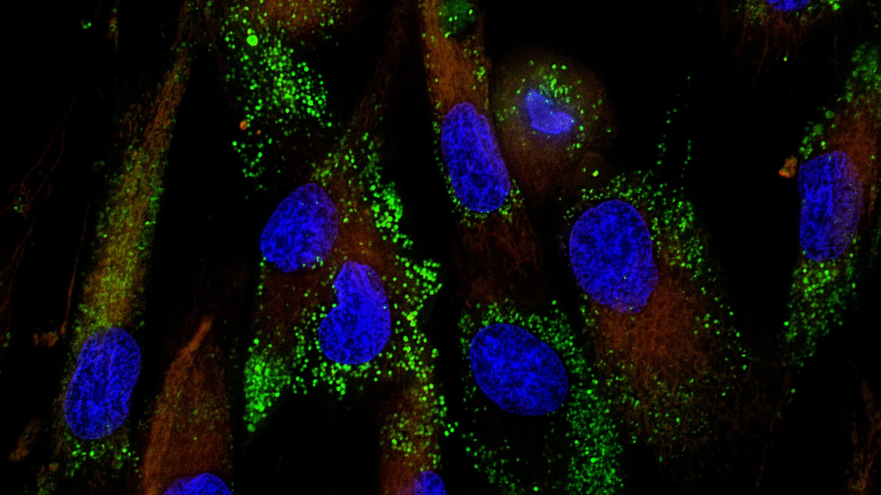
Imaging nucleic acids (DNA, RNA) is possible but has not been met with as much success as protein imaging. Nucleic acid imaging of fixed cells is done through a process called fluorescence in situ hybridization (FISH). FISH is analogous to IF/IHC except instead of antibodies, oligos are used to bind to nucleic acids and amplification is necessary to visualize targets due to the low abundance compared to proteins.
However, the ability to image nucleic acids in real-time has been more challenging due to spatial and size constraints. Scientists developed a method to fluorescently tag DNA-binding proteins. However, this method is heavily limited by targeting sequences to certain genomic regions. Further, this technique cannot bind non-repetitive sequences, which accounts for a large percentage of the genome.
The need to see nucleic acids in real time is becoming more apparent since scientists now believe cellular dynamics are much more complicated than initially thought. While the aforementioned techniques are still being used, their inherent limitations are not providing scientists with an accurate story. Therefore, there needed to be a way to visualize proteins and nucleic acids in real time and space to understand how three-dimensional genomic architecture plays a vital role in regulating gene expression and cell behavior. This is where the guiding and DNA binding power of CRISPR comes in to help.
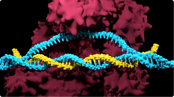
CRISPR-Cas9 based imaging techniques allow scientists to track genomic loci of interest in real time. Cas9 is modified to contain an imaging probe (i.e., fluorescent protein) and introduced into cells along with a guide RNA. Following delivery, the guide RNA directs the labeled Cas9 to the site of interest and enables imaging of that particular DNA target.
The first description of CRISPR-Cas9 as an imaging tool was published in 2013 by the groups of Stanley Qi and Bo Huang. In this study, researchers used an inactive Cas9 (no cutting activity) tagged to an enhanced green fluorescent protein (EGFP) and a guide RNA. EGFP is commonly used to visualize cells because it produces a robust bright green color that is easily detected. The modified Cas9 and guide RNA were introduced into cells and scientists were able to detect EGFP inside the cells.
Researchers could successfully use this new method to visualize a number of sequences. This seminal work has enhanced the applications of CRISPR imaging, providing scientists with a new method of visualizing cellular dynamics at a high resolution.
Now that we have the tools, what are scientists using CRISPR imaging for? Since this is a fairly new technology, research efforts surrounding CRISPR imaging fall under two main categories: chromatin remodeling and telomere health.
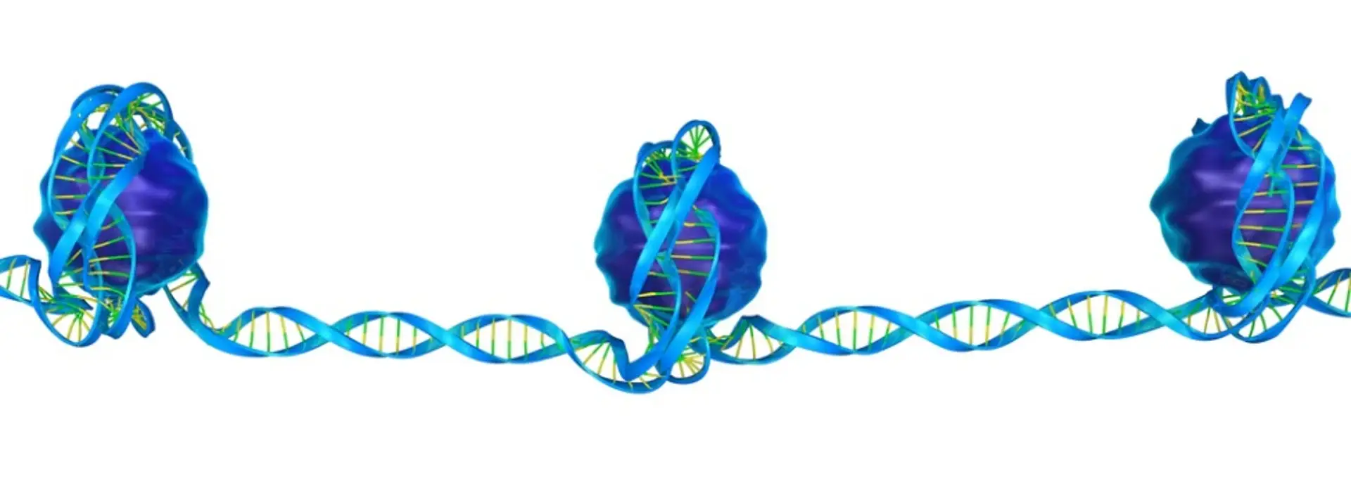
Chromatin remodeling is the rearrangement or modification of chromatin that is integral for facilitating interaction between DNA and transcription factors, the two key players of regulating gene expression. Since the involvement of gene expression is ubiquitous in biological processes, understanding the mechanisms underlying chromatin remodeling are vital.
By targeting sequences known to be involved in chromatin remodeling, scientists can mark them with fluorescently labeled Cas9, and then track the dynamics with real-time imaging. For example, we can watch the chromosomal dynamics of MUC4 as the cell goes through the phases of cell division. Additionally, scientists are using CRISPR imaging methods to visualize chromatin dynamics in yeast cells in response to nutrient starvation.
Telomeres are regions of repetitive sequences at the ends of chromosomes that protect the chromosome from deteriorating, and thereby are critical to genomic health. Reduction of telomere length is often seen in aging-related diseases and is also associated with many cancer types. Therefore understanding telomere dynamics, and how scientists can prevent telomere shortening, is a crucial aspect of addressing human health.
As mentioned above, this CRISPR imaging technique allowed imaging telomeres. Researchers were able to visualize telomere expressing cells by fluorescent dots (or puncta) at levels comparable to telomere-specific FISH. Furthermore, they were also able to monitor telomere length and movement. Additionally, visualizing of telomere dynamics in plants have also been reported.
Although great progress has been made in the CRISPR imaging field, several challenges exist. In order to improve this technology, scientists are currently trying to find solutions to the challenges described in this section.
All imaging techniques suffer from the delicate balance between the signal-to-background ratio. The signal-to-background ratio refers to the ratio of true fluorescence intensity against the background intensity or non-specific signal. Maximizing the signal-to-background ratio is important for knowing if what you are seeing is truly Cas9 bound to your target site of interest.
The main way that scientists are trying to address this is by optimizing their transfection conditions. One of the main reasons for background noise is the presence of unbound or free-floating fluorescently-labeled reagents. Even if it doesn’t bind its DNA target, the labeled Cas9 will still fluoresce and be detectable. If the transfection is optimized, the amount of unbound Cas9 and sgRNA can be minimized, decreasing the likelihood of this problem.
Imaging DNA using tagged DNA-binding proteins has been restricted to repetitive sequences, which only accounts for a small percentage of the genome. Visualizing non-repetitive sequences was not possible with this previous method. However, the target specificity of Cas9 solves this problem.
In order to target non-repetitive sequences, scientists needed a way to deliver multiple sgRNAs targeting adjacent sites. Scientists developed ways to clone multiple sgRNAs into one plasmid for delivery. Two methods have been described with this principle in mind. The first one is chimeric array of gRNA oligonucleotides (CARGO) which, when coupled with Cas9, results in the delivery of multiple gRNAs into cells. Golden Gate Assembly is another transfection method aimed to increase efficiency.
Despite these efforts, the results are not perfect. Scientists are now exploring other potential ways to address this issue, including linking sgRNAs by tRNA through a process termed (Po)STAC (Polycistronic SunTAg modified CRISPR), re-engineering sgRNAs to contain motifs that improve targeting, or possibly using different orthologs of Cas9 to ensure cutting by multiple sgRNAs.
Cas9 (specifically SpCas9) is the most commonly used Cas variant used for CRISPR imaging. It recognizes a 3-base pair PAM sequence that increases the number of target sites but comes at the cost of increased off-target effects. This can lead to false positives in imaging (i.e. labeling the wrong region).
Scientists are addressing this challenge by using other variants of Cas9 that have longer PAM sequence requirements. This limits the number of usable target sites, however, increases the specificity.
Even though challenges still remain, CRISPR as an imaging tool is already proving to be highly useful for understanding cellular dynamics. The CRISPR imaging field is just emerging, so be sure to look out for new applications and discoveries in the future.
This protocol describes the use of limiting dilution to isolate single cells (clones) from a CRISPR edited population, the expansion of clonal populations, and screening to identify clones with the...
Thank you for choosing SpCas9 nuclease protein and gRNAs for your CRISPR experiment! SpCas9 has been used for diverse genome editing applications, making it the gold standard in CRISPR gene...
Learn more about clinical applications of CRISPR and how Synthego can support you from early-phase research, through process development, and into the clinic, highlighting our new sgRNA GMP manufacturing capabilities.